![]()
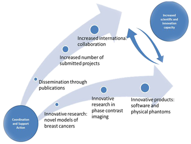
L'obiettivo di SYRMA-CT è di mantenere la leadership italiana nella mammografia in contrasto di fase estendendo il programma clinico della linea SYRMEP alla tomografia della mammella, per ottenere immagini CT mammografiche che sfruttino al meglio gli effetti di fase che non sono osservabili con gli attuali sistemi CBBCT. A questo scopo le competenze di eccellenza maturate nell’INFN nell’ambito delle attività di CSN5 confluiscono nel progetto per permettere il raggiungimento degli obiettivi in tempi brevi e con alte probabilità di successo.
Aspetto critico per il successo del progetto sarà l'ottimizzazione dosimetrica che implica una opportuna scelta dell'energia (nel range 20-38 keV), della risoluzione spaziale del detector (risoluzione spaziale di base 65 micron), della modalita’ e dei parametri di acquisizione tomografica. In questo campo le competenze accumulate nei progetti di cone-beam CT dei gruppi di Napoli e Bologna saranno ampiamente utilizzate.
Synchrotron Radiation Rotational RadioTherapy for Breast Cancer
Project Acronym: SR3T
Starting date: 01/01/2018
Duration in Months: 36
Principal Investigator: Giovanni Mettivier, PhD
Objective: To propose and investigate an innovative therapeutic approach for breast cancer therapy which combines kilovoltage external beam rotational radiotherapy using synchrotron radiation, the use of contrast and radiosensitizing agents, image guidance via pre-treatment and within-treatment computed tomography scans for precise tumor localization and patient repositioning, showing its potential with respect to currently available conventional and experimental radiotherapy techniques for breast cancer.
Abstract
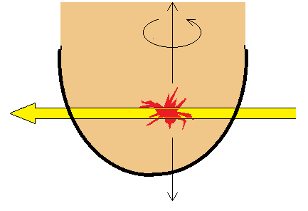
Radiotherapy is normally delivered using high-energy MeV photon beams produced by clinical accelerators. Radiation therapy of breast cancer may benefit from recent experimental investigations in tumor radiotherapy with kilovoltage photon beams produced by orthovoltage X-ray tubes. We propose to adopt monoenergetic or polyenergetic beams at photon energies below @ 200 keV down to @ 60 keV for breast cancer rotational radiotherapy, either fully opened to irradiate the whole breast, or suitably collimated to irradiate at least part of the tumor volume, then scanned across that volume in a fully tomographic rotate-and-translate geometry. The source for such an experimental technique is a medium-energy synchrotron radiation. In this new technique, termed synchrotron radiation rotational radiotherapy (SR3T), the patient is in prone position on a bed containing a hole through which the uncompressed breast hangs inside a holder. The bed is rotated over 360 deg around a vertical axis centered at the tumor site, with the beam suitably collimated in the horizontal and vertical directions, so as to irradiate, in the horizontal plane, the whole breast or just a thin tumor slice. The bed is then translated vertically and another slice of the target volume is irradiated in a successive full rotation: after a suitable number of rotations (with possibly laterally displaced vertical axes of rotation), all the target volume may receive the planned dose. Preliminary Monte Carlo investigations and experimental validations recently performed by the proponents indicated that ─ by the principle of rotational summation of dose at the axis of rotation ─ the almost exponentially decreasing trend of the absorbed dose with radial depth in the medium characteristic of the single-irradiation angle, turns into a dose distribution peaked at the axis of rotation, where the target volume is located. This produces an inversion of the percentage depth dose curve, with less dose at the organ surface than at the tumor site, assuring similar values of the skin sparing as with conventional accelerator based breast radiotherapy with two 180-deg angled beams tangential to the chest wall, due to the presence of the build-up region for megavoltage beams (15 mm maximum dose depth at 6 MV in water). This three-year project proposes to carry out Geant4 Monte Carlo simulations and experimental validations in breast phantoms at synchrotron radiation facilities, for assessing the merits of the SR3T technique, in terms of 3D dose distributions and tumor-to-skin dose ratio, cell culture studies for cell killing action at kilovoltage photon energies, possible use of iodinated contrast agents and metal nanoparticles as radiosensitizing agents, optimal energy selection, full target volume coverage, identification of basic parameters for a future treatment planning system at synchrotron radiation facilities.
Project Highlights:
- Breast radiotherapy with kilovoltage X-ray beams
- dose enhanced radiotherapy with iodinated agent
- Rotational irradiation in full tomotherapy for skin sparing without buildup
- Online image guided breast CT
- Collimated beam or pencil beam scanning for dose painting
- Contrast enhanced CT
 |
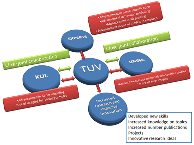
MaXIMA Project -Three dimensional breast cancer models for X-ray Imaging research- The development of realistic three-dimensional computational and physical models of breast tumours with irregular shapes and their availability is a powerful instrument in the hands of engineers, physicians and physicists to be used in the development of new technologies for precise definition of the cancer boundaries. Scientists from the biomedical engineering unit at the Technical University of Varna work in this area, both on modelling and simulation of computational breast phantoms and x-ray breast imaging techniques. To advance in their research aims and therefore to raise the profile of their researchers and S&T capacity of the host organisation, collaborations with top research institutions from complementary areas of expertise are a requirement.
|
 |
The main objective of MAXIMA action is to increase the research and innovation capacity of the host organisation in the field of computational modelling of breast tumours (including cancers with irregular shape) and their use in studies of advanced x-ray breast imaging techniques such as breast tomosynthesis and phase contrast imaging. For this purpose, the project will establish a close and sustainable collaboration platform, in the form of a network of three scientific groups working in the specific domain of modelling and simulations dedicated for studies of x-ray breast imaging techniques. |
|
Partners: Technical University of Varna Katholieke University of Leuven University of Naples, Federico II
|
|